CELL MODEL DATASHEET
Diverse-donor endothelial cells for vascular, barrier and liver co-culture models

Cytochroma human iPSC-derived endothelial cells are cryopreserved endothelial cells differentiated from an ancestrally and genetically diverse, HLA-typed induced pluripotent stem cell library. Endothelium controls the barrier between blood and tissue, so it governs how much of a compound reaches its target and how immune cells traffic into an inflamed organ. Supplying endothelial cells from the same genotyped donors as Cytochroma hepatocytes and Kupffer cells allows vascularised and sinusoid-like liver models to be built in one consistent genetic background.
8 / 18
donor lines fully HLA-typed
71
distinct HLA alleles across the panel
1:1
female to male donor balance
3
Mapped to 3 diverse ancestry groups
Why this model
IComplete the microenvironment
Endothelial cells add the vascular compartment to hepatic co-cultures and microtissues, alongside hepatocytes, Kupffer cells and hepatic stellate cells from the same donor library.
Genotype-matched or deliberately mismatched
With HLA typing at two-field resolution across eight lines, endothelial and immune compartments can be matched or mismatched on purpose for allogeneic and vascular immune studies.
Renewable supply
Differentiated to order from a defined iPSC library rather than sourced from scarce primary donor tissue.
specifications
Cell type
Human iPSC-derived endothelial cells
Donor panel
8 of 18 lines fully HLA-typed at two-field resolution; 71 distinct HLA alleles; 1:1 female to male
Viable cells per vial
Greater than or equal to 1 x 10⁶ (1.63 x 10⁶ recorded for lot MAN019-31E)Post-thaw viability
Post-thaw viability
≥70% by automated trypan blue exclusion; 96.0% recorded for lot MAN019-31E
Format
Cryopreserved, 1.0 mL per vial; store at or below -145 °C; 12 months from receipt
Identity markers
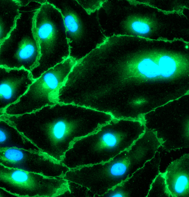
CD31 (PECAM-1) detected by immunofluorescence at QC4; polygonal endothelial morphology confirmed by microscopy. CD144 (VE-cadherin) and CD34 also detected by immunofluorescence.
Functional characterisation
Dil-Ac-LDL uptake demonstrated at QC4 (10 µg/mL, 1 hour, 37 °C). Forms capillary-like networks in three-dimensional spheroid culture, with continuous CD144 junctional staining consistent with adherens-junction formation.
Documentation
Certificate of Analysis with every lot: viability, viable recovery, mycoplasma (MycoAlert, ratio less than 1.0), bacterial and fungal sterility, adherence at 24 hours, morphology, Dil-Ac-LDL uptake and CD31 staining, with brightfield and fluorescence images.
Sterility and mycoplasma
Mycoplasma negative by MycoAlert activity assay (ratio less than 1.0); bacteria and fungi negative by visual assessment at QC4
Manufactured in
Roslin Innovation Centre, Edinburgh, United Kingdom

Applications
Vascularised liver microtissue and organ-on-chip models
Barrier integrity and permeability studies
Endothelial activation and immune cell adhesion assays
Angiogenesis and tube formation assays
Donor-stratified vascular toxicity screening
Co-culture with iPSC-derived hepatocytes, Kupffer cells and stellate cells
Donor diversity and HLA typing
Every Cytochroma cell model is differentiated from an induced pluripotent stem cell library built deliberately for population-level variability rather than convenience. 8 of 18 of 18 iPSC donor lines fully HLA-typed at two-field resolution. 71 distinct HLA alleles across the panel. 1:1 female to male donor balance. European, Native American and African ancestry represented. Pairwise HLA mismatch scores of 7 to 10 out of 10 between almost every donor pair, across HLA-A, B, C, DRB1 and DQB1.
Typing covers the classical class I loci HLA-A, HLA-B and HLA-C and the class II loci DRB1, DRB3/4/5, DQA1, DQB1, DPA1 and DPB1 at two-field resolution. Full multi-field typing is available on request. Because almost every donor pair scores 7 to 10 out of 10 on pairwise allele-level mismatch, donor-mismatched co-culture, allogeneic response and HLA-restriction studies can be set up off the shelf.
Evidence and validation
PShared donor library
Endothelial cells draw on the same HLA-typed, ancestrally diverseiPSC library documented for the Cytochroma hepatic range.
Frequently asked questions
What are iPSC-derived endothelial cells used for?
Are the endothelial donor lines HLA-typed?
Can endothelial cells be supplied from the same donor as the hepatocytes?
How do I order?
Request a quote or technical discussion
Tell us your assay format, donor requirements, and timelines. Our scientific team will help select the right donor panel and model configuration.